
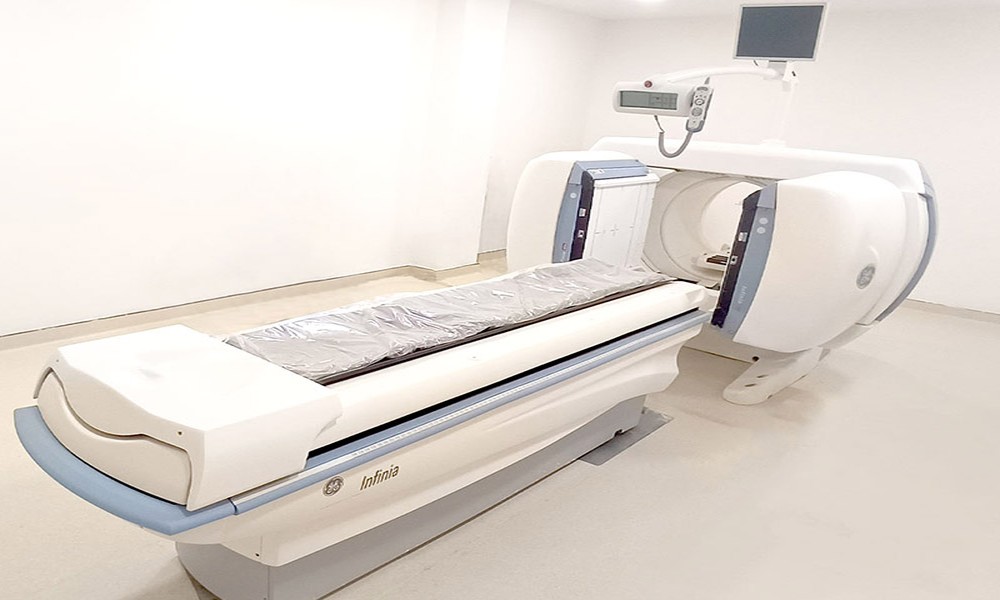
Gamma camera is a nuclear medicine imaging modality which provides vital details regarding the physiological functions of organs and tissues. Unlike other scans that focus on structure, it looks at the activity inside the body. It consists of components such as collimators, scintillators, photomultiplier tubes, and advanced imaging software. You might also hear it called a scintillaiton camera.
Doctors commonly use gamma camera scan to study organs such as the heart, lungs, thyroid, liver, kidneys, and brain. It plays an important role across multiple specialities such cardiology, oncology, neurology, endocrinology, and orthopedics. It is also useful in analysing how far the disease may have spread (in certain cancers affecting organs or bones) and in tracking how well a treatment is working over time.
How does a gamma camera operate?
The process begins by using a small amount of radiotracer, most often technetium-99m, which is injected into a vein. This tracer travels through the body and settles in organs or tissues that need to be examined.
As it moves, it emits tiny energy signals known as gamma rays. These photons escape the patient’s body and are picked up by the gamma camera. A part called collimator helps filter out unwanted signals so only useful ones are captured.
Inside the camera, a special crystal converts these signals into tiny flashes of light, which are then amplified and turned into images that show how the tracer is distributed in two dimension. Due to the fact that the specialised crystal emits tiny flashes of light – it is also called a scintillation camera.
In more advanced scans like SPECT, the camera slowly revolves around the body to capture cross-sectional or volumetric images. This means SPECT imaging with a CT scan can deliver 3D images. This helps the clinicians to clearly understand how the organs are functioning, even before the structure changes are visible. Depending on the type of scan, the process can take anywhere between 40 mintues and a couple of hours.
If you’re based in Delhi-NCR or Mumbai, a quick online search can help you find out gamma camera scan cost in your area.
Benefits
1, While the traditional scans show the images on a structural level, a gamma camera can tell how the heart is functioning or how the blood is perfusing a part of the brain.
2. Given that gamma camera can show the functional changes, the clinicians can pick up the illnesswa in their early phase. This allows for better patient outcomes.
3. The gamma camera provides the flexibility of scanning a wide range of organs such as the liver, the kidneys, bones, thyroid, the heart (myocardial perfusion imaging) and brain (SPECT brain). They are often used for bones infections and cancer.
4. When combined with a CT scan, you get anatomical context plus functional data.
5. This can can also be used to evaluate organ function post-procedure, monitor therapies and plan surgical interventions.
Preparations
Before going for the scan, the patient needs to bring the doctor’s prescription and any previous reports. The patient will be required to take off some or all of the clothing and wear a hospital gown, remove any metal objects such as trouser braces, jewellery and belts.
In most cases, no dietary restriction is required for the procedure, so you can eat and drink normally. For this scan, a small amount of radiotracer will be injected through the arm which will be taken up by specific part of the body. The emitted radiation gets detected by the camera to deliver an image.
While images for some scans can be taken immediately, for others there could be a waiting period of upto 3 hours. Once the scan the completed, the patient can leave the diagnostic facility immediately.
Disclaimer: For personalised advice and further information, always consult your doctor or qualified healthcare professional.